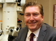
David R. Hinton, MD, FARVO
Professor of Pathology, Neurological Surgery, Ophthalmology; Associate Dean for Vision Science; Vice Chair for Research, Exam Committee Facilitator; Director, Dean’s Research Scholars Program
Dr. David Hinton’s research is focused on developing novel therapies for age-related macular degeneration (AMD) – the leading cause of irreversible blindness in the elderly. He is approaching this goal in two main ways: (1) restorative cell therapy to replace cells already damaged, and (2) neuroprotection to prevent the retinal degeneration. A primary site of pathology in AMD is the retinal pigmented epithelial cell (RPE). Dr. Hinton has been studying the basic biology of the RPE and its response to injury for over 20 years. He established the importance of RPE-derived growth factors in AMD and other retinal disorders including vascular endothelial growth factor (VEGF), connective tissue growth factor (CTGF) and hepatocyte growth factor (HGF). He has developed a well-known in vitro model of polarized RPE cell monolayer culture and has established several in vivo models that recapitulate several features of the pathophysiology of AMD. He has developed (in collaboration with Dr. Mark Humayun at USC and Dr. Dennis Clegg at UC Santa Barbara) a novel cell therapy approach to the late, dry form of AMD. Embryonic stem cells are differentiated into RPE and then grown as a polarized monolayer for subretinal implantation in patients. With extensive funding from the California Institute for Regenerative Medicine (CIRM), this team is completing preclinical studies demonstrating safety and efficacy of the approach. They are planning to submit an investigational new drug (IND) application to the FDA this fall and have already obtained funding to begin a Phase 1 safety trial in AMD patients in 2015.
Dr. Hinton has also established a program to develop drugs that would protect RPE cells and light sensitive photoreceptors from damage. Such drugs could be used as a preventative measure or in conjunction with cellular therapy. Hinton is particularly interested in the small heat shock protein alphaB-crystallin. He has shown that it strongly protects RPE from oxidative and endoplasmic reticulum stress. He then showed that alphaB-crystallin is released from the apical surface of RPE cells in exosomes where it can protect adjacent cells. He is leading a NIH-funded effort with Dr. Ram Kannan (Doheny Eye Institute) and Dr. Andrew MacKay (USC School of Pharmacy) to develop small peptides of alphaB-crystallin into a therapeutic for patients with AMD or other retinal degenerations. Dr. Hinton has published almost 300 peer-reviewed papers and is a frequent speaker at national and international meetings to present his work. Dr. Hinton was named a “Master Teacher” at KSOM in 2008.
We asked Dr. Hinton to answer a few questions about his decision to study macular degeneration, his thoughts on future research in this area, and to tell us about the Tissue Imaging Core he oversees.
What personal or professional experiences influenced your decision to study AMD?
I was trained as a board-certified Neuropathologist whose main role was to evaluate tissue samples obtained by Neurosurgeons at the time of brain surgery. While this was clinically fulfilling, I was very interested in establishing a research program that could determine basic mechanisms of neurologic disease and eventually develop novel therapies. I learned that the retina, the neural tissue at the back of the eye, represented an approachable part of the nervous system in which mechanisms of disease could be studied and evaluated more easily. I initially studied tumors of the retina (retinoblastoma) as a Neuropathology resident, and then as a postdoctoral fellow I studied the development of the retina in the research labs of Dr. Seymour Benzer (at Caltech) and Dr. Carol Miller (at USC). Once I was recruited to USC, I was strongly influenced by my research mentor, Dr. Stephen Ryan, who introduced me to the tremendous unmet need for preventing and treating the blindness caused by AMD. It was at that point that I focused my efforts on the RPE cell, which at the time was being recognized as the primary site of pathology in AMD. After meeting many people with AMD I realized that I had the opportunity to study the basic biology of the RPE and through these efforts developed novel therapies that could potentially treat or even prevent the blindness caused by AMD.
What would you say is the most significant advance in this field as contributed by your own research?
I have been a leader in establishing the critical role of the RPE in the pathogenesis of AMD. I was the first to show expression of Vascular Endothelial Growth Factor (VEGF) in RPE from AMD patients with the late, wet form of the disease in 1996. Since that time, anti-VEGF therapies have revolutionized the therapy of these patients. I have promoted the concept of RPE function as a polarized monolayer, and have established in vitro models of RPE monolayer formation. I initiated the original studies at USC to differentiate embryonic stem cells into RPE and established a collaboration with Dr. Mark Humayun to form a team of investigators that have gone on to develop a cellular therapy for AMD that may soon be in clinical trials. I am developing novel neuroprotective therapies that could help to prevent the progression of retinal degeneration in patients with AMD.
What other areas of research have you collaborated with and how have these collaborations advanced or influenced your own research?
When I was recruited to USC, I was very fortunate to continue collaboration with my postdoc mentor, Dr. Carol Miller. My first paper at USC was a first author publication in the New England Journal of Medicine (coauthored by Dr. Carol Miller and Dr. Alfredo Sadun). In this paper we described for the first time a retinal degeneration in patients with Alzheimer’s disease. Since that time, over 200 publications have been written that further evaluate this change and its potential in using the eye as a “window to the brain.” In exciting recent studies, we are continuing this work using novel technologies with collaborators at Cedars Sinai.
I was also very fortunate to collaborate with Dr. Florence Hofman in the department of Pathology. Dr. Hofman introduced me to the field of neuroimmunology and among our studies was a paper in Journal of Experimental Medicine that demonstrated the expression of tumor necrosis factor in the brains of patients with multiple sclerosis. I went on to establish collaborations with Drs. Steve Stohlman and Conni Bergmann at Cleveland Clinic where we have been funded by NIH to study the role of immunoregulatory molecules in animal models of demyelination for over 20 years. The expertise I developed in these projects has been very influential in developing concepts about the role of inflammation in AMD.
A third major area of collaboration has been with Dr. J. Ambati at the University of Kentucky. This collaboration developed as a result of my expertise in the culture and evaluation of human RPE. Over the past five years I have co-authored seminal papers with him on the basic pathogenesis of AMD in PNAS, Cell, Nature, Nature Medicine, and the New England Journal of Medicine.
You also oversee the Cell and Tissue Imaging Core. Could you describe some of the resources of this core and how other USC investigators can benefit from its use?
The Cell and Tissue Imaging Core Facility is operated under my direction and supervised by Mr. Ernesto Barron. It provides support for research projects requiring specialized microscopic imaging. The facility is currently supported by a National Cancer Institute (NCI) Core grant to the Norris Cancer Center. The Cell and Tissue Imaging Core is available by appointment, 24 hours/day, seven days per week. The primary function of the Core is to provide assistance and training in the operation of equipment for laser scanning confocal microscopy, multiphoton laser scanning confocal microscopy (Zeiss LSM510 and Zeiss LSM710), spinning disk confocal microscopy (PerkinElmer Ultraviewer spinning disk confocal), transmission electron microscopy (JEOL JEM 2100 LaB6), scanning electron microscopy (JEOL JSM/6390LV), Laser Capture Microdissection Microscopy (Zeiss PALM System), Digital Scan Scope Microscopy (Aperio Scan Scope), fluorescence microscopy, quantitation of immunocytochemical procedures (Leica and Olympus Microscopes) and fluorescence and brightfield microdissection and microcapture (Zeiss PALM System). The Core offers support for specimen preparation, thin sectioning techniques, embedding procedures, cryosectioning techniques, photography, digital photomicroscopy and photomicrography and computer-aided graphics. Further information is available at https://research.usc.edu/
What do you think will be the next big advances in your field and what do you think KSOM can do to position itself to be a leader in this field?
There are going to be rapid advances in the prevention and treatment of blindness, including regenerative cellular therapy (not only of the RPE but also the photoreceptors and potentially the whole retina), and neuroprotective therapies (to prevent retinal degeneration). Dr. Mark Humayun has been a leader in the development of the retinal prosthesis that has regulatory approval for the treatment of patients with retinitis pigmentosa and has brought functional vision to those that were previously blind. The new field of optogenetics, in which neurons which have been genetically sensitized to light can be controlled by light as an alternate approach to bring vision to blind patients is under investigation at USC. USC is also at the leading edge in development of high resolution imaging of the retina and central visual pathways that will allow better understanding of retinal disease and its response to therapy. The USC Department of Ophthalmology and the USC Eye Institute will be the focal points for these developments with highly collaborative interactions throughout the university.